



















ABG Interpretation Made Easy: 4-Step Guide for Students
ABG interpretation is one of the most challenging yet essential clinical skills for medical students and healthcare professionals. Interpreting arterial blood gas (ABG) results means connecting numbers with real clinical decisions, which many learners find difficult.
This guide explains ABG analysis step by step combining normal values, interpretation logic, and practical examples to help you interpret ABGs confidently in exams and patient care. ABG analysis measures oxygen (PaO2), carbon dioxide (PaCO2), and pH, assessing oxygenation, ventilation, and acid-base balance, and is more accurate than non-invasive methods like pulse oximetry.
Clinical Importance and Indications
ABG analysis plays a central role in evaluating and managing many conditions:
- Respiratory failure (acute or chronic)
- ARDS, sepsis, or shock
- Diabetic ketoacidosis and renal tubular acidosis
- Heart failure, cardiac arrest, and asthma
- Inborn errors of metabolism
Bicarbonate (HCO3) is usually calculated from ABG values, and small differences may exist between calculated and measured values, especially in critically ill patients.
Technical Challenges
Arterial sampling can be difficult in patients with shock, obesity, weak pulses, or positioning limitations. Ultrasound-guided puncture significantly improves success rates and reduces complications from repeated attempts. Frequent ABG monitoring is common in shock (to assess lactic acidosis and respiratory compensation) and COPD exacerbation (to assess respiratory acidosis and CO2 retention).
Practical Steps for Obtaining an ABG Sample
- Sampling method: needle puncture for occasional tests; an indwelling catheter (with written consent) if frequent draws are needed. If sampling more than four times in 24 hours, rotate sites or consider a catheter.
- Site selection: radial, brachial, femoral, axillary, or dorsalis pedis. The radial artery is most used for accessibility and comfort.
- Ensure collateral circulation: for small arteries (radial, dorsalis pedis), perform the Allen / Modified Allen test; color should return within about 10 seconds. Large arteries usually do not require collateral testing.
- Equipment: heparinized syringe, needle and cover, ice bag to preserve the sample, and local anesthetic (e.g., lidocaine) if needed.
- Puncture details: radial at ~30 degrees palm-up; brachial medial to the biceps tendon at ~30 degrees; femoral below the inguinal ligament midpoint at 90 degrees; dorsalis pedis at ~30 degrees.
Before starting ABG interpretation, it is important to understand the normal reference values for pH, PaCO2, PaO2, HCO3, and oxygen saturation.

Normal ABG Reference Values
- pH: 7.35–7.45 (<7.35 acidemia, >7.45 alkalemia)
- PaCO2: 35–45 mmHg (>45 respiratory acidosis, <35 respiratory alkalosis)
- PaO2: 80–100 mmHg (<80 hypoxemia)
- HCO3: 22–26 mEq/L (<22 metabolic acidosis, >26 metabolic alkalosis)
- Base excess: -2 to +2 mEq/L
- SaO2: 95–100% (<95% low saturation)
- Anion gap: 8–12 mEq/L (>12 high anion gap metabolic acidosis)
Normal ranges vary slightly by laboratory, altitude, and clinical condition; always interpret in clinical context.
A systematic approach makes ABG interpretation easier, especially when identifying the primary disorder and checking whether compensation is present.
ABG Interpretation: The 4-Step Method
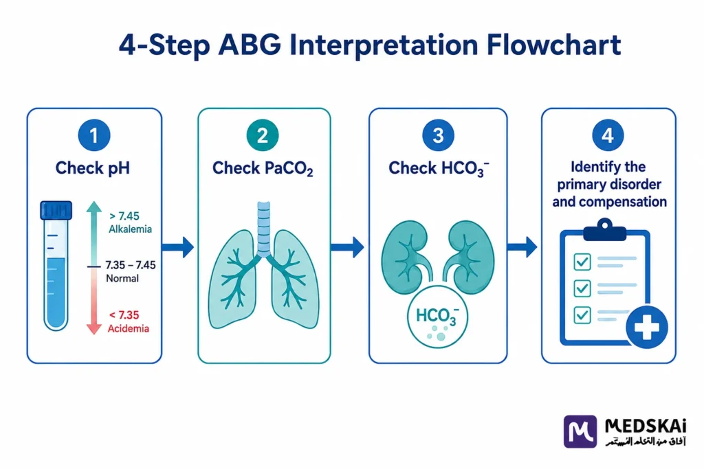
- Assess the pH: <7.35 acidosis, >7.45 alkalosis; a normal pH may indicate full compensation or a mixed disorder.
- Identify the primary disorder: if PaCO2 is abnormal and moves opposite to pH, the disorder is respiratory; if HCO3 is abnormal and moves in the same direction as pH, it is metabolic.
- Assess compensation: metabolic acidosis lowers PaCO2 and metabolic alkalosis raises it; chronic respiratory acidosis raises HCO3 and chronic respiratory alkalosis lowers it (metabolic compensation takes 3–5 days).
- Calculate the anion gap in metabolic acidosis: Anion Gap = Sodium – (Chloride + Bicarbonate); normal 8–12. High (>12) suggests unmeasured acids (DKA, lactic acidosis); normal suggests bicarbonate loss (e.g., severe diarrhea).
A normal pH does not always mean a normal acid-base state — always consider mixed disorders and assess compensation.
Common ABG Interpretation Mistakes
Frequent errors include relying on pH alone, ignoring compensation, misinterpreting a normal pH in mixed disorders, and failing to calculate the anion gap in metabolic acidosis. For example, a normal pH with abnormal PaCO2 and HCO3 often indicates a mixed disorder rather than normal physiology.
Complications and Contraindications
Sampling complications include hematoma, bleeding, infection, vasospasm, and rare nerve injury. Misinterpretation can delay diagnosis or lead to inappropriate treatment. Severe acidosis may cause arrhythmias and reduced response to inotropes; severe alkalosis may cause cerebral vasospasm, seizures, and hypokalemia.
Relative contraindications include deficient collateral circulation, overlying skin infection, coagulopathy or anticoagulation (sample only if necessary), and severe vascular disease making access difficult.
Frequently Asked Questions
What does ABG analysis tell us about a patient’s condition?
It shows how well the lungs are working, how much oxygen reaches the blood, and whether acid-base balance is normal making it key for assessing respiratory and metabolic status.
What are the four steps of ABG interpretation?
Assess the pH, identify the primary disorder (respiratory vs metabolic), evaluate compensation, and calculate the anion gap in metabolic acidosis.
What is the anion gap and why does it matter?
Anion Gap = Sodium – (Chloride + Bicarbonate), normally 8–12 mEq/L. A high value suggests unmeasured acids (e.g., DKA, lactic acidosis); a normal value suggests bicarbonate loss.
Why use ultrasound for arterial sampling?
In patients with shock, obesity, or weak pulses, ultrasound guidance improves success rates and reduces complications from repeated punctures.
Conclusion
ABG interpretation remains a vital diagnostic tool, especially in critically ill patients. Correct sampling technique combined with systematic interpretation maximizes its value.
Always interpret ABG results within the patient’s complete clinical picture rather than in isolation.
This article is for educational purposes only and does not replace consultation with a qualified medical specialist.
Move from memorizing ABG values to truly understanding them — explore MedSkAI’s step-by-step ABG course with real ICU and ER cases. Courses – MedSkAI
External References
For deeper, source-based reading on ABG interpretation, see StatPearls: Arterial Blood Gas (NCBI Bookshelf) and LITFL: ABG Interpretation.
- Castro, D., Patil, S. M., Zubair, M., & Keenaghan, M. (2024).
- Arterial blood gas. In StatPearls. StatPearls Publishing. Rogers, J., & Cadogan, M. (2025, March 12).
- Blood gas interpretation. Life in the Fast Lane. Kaufman, D. A. (n.d.). Interpretation of arterial blood gases (ABGs). American Thoracic Society.