


Course Overview:
Magnetic Resonance Imaging (MRI) is one of the most advanced and powerful imaging modalities in modern medicine, providing exceptional soft tissue contrast without the use of ionizing radiation. A deep understanding of MRI requires not only knowledge of imaging protocols, but also a solid foundation in physics, signal generation, and image formation.
This comprehensive course is designed to provide a structured learning pathway that guides participants from the fundamental principles of MRI physics, through signal formation and image contrast, to understanding MRI sequences, parameters, and clinical applications. The course integrates theoretical concepts with practical insights to help learners confidently interpret MRI images and understand how imaging decisions affect diagnostic outcomes.
The program is organized progressively, starting from the basic concepts of hydrogen atoms and magnetic fields, moving through RF excitation and relaxation mechanisms, and advancing toward image reconstruction, sequences, artifacts, and safety considerations in clinical MRI practice.
What you’ll learn
This course provides a complete and structured understanding of MRI, starting from the physical principles of signal generation to advanced imaging techniques and clinical applications.
-
- Fundamentals of MRI physics including hydrogen behavior, magnetic field (B₀), and proton alignment
- Principles of precession, Larmor frequency, and RF excitation
- T1 and T2 relaxation mechanisms and their impact on image contrast
- MRI system components, including magnet, gradients, RF system, and computer system
- Spatial encoding techniques (slice selection, frequency encoding, phase encoding) and k-space fundamentals
- MRI parameters such as TR, TE, flip angle, and their role in image optimization
- MRI contrast types (T1, T2, Proton Density) and clinical relevance
- Common MRI sequences including Spin Echo, Gradient Echo, FLAIR, and STIR
- MRI artifacts and techniques for improving image quality
- MRI safety principles, hazard management, and patient handling
Target Audience
- Radiology and medical imaging students
- MRI trainees and newly graduated radiographers
- Radiologic technologists seeking to strengthen MRI fundamentals
- Healthcare professionals interested in understanding MRI imaging
- Anyone aiming to build a strong foundation in MRI physics and clinical application
- 7 Sections
- 10 Lessons
- 365 Days
- Introduction1
- MRI Scientific principle5
- 2.1MRI Scientific principle29 Minutes
- 2.2Quiz115 Minutes15 Questions
- 2.3MRI Scientific principle (T1 and T2 relaxation mechanisms and their impact on image contrast)35 Minutes
- 2.4Spatial encoding techniques (slice selection, frequency encoding, phase encoding) and k-space fundamentals35 Minutes
- 2.5Quiz215 Minutes15 Questions
- MRI Sequences1
- MRI parameters2
- MRI system components1
- MRI Hardware & Patient safty2
- MRI artifacts and techniques for improving image quality3

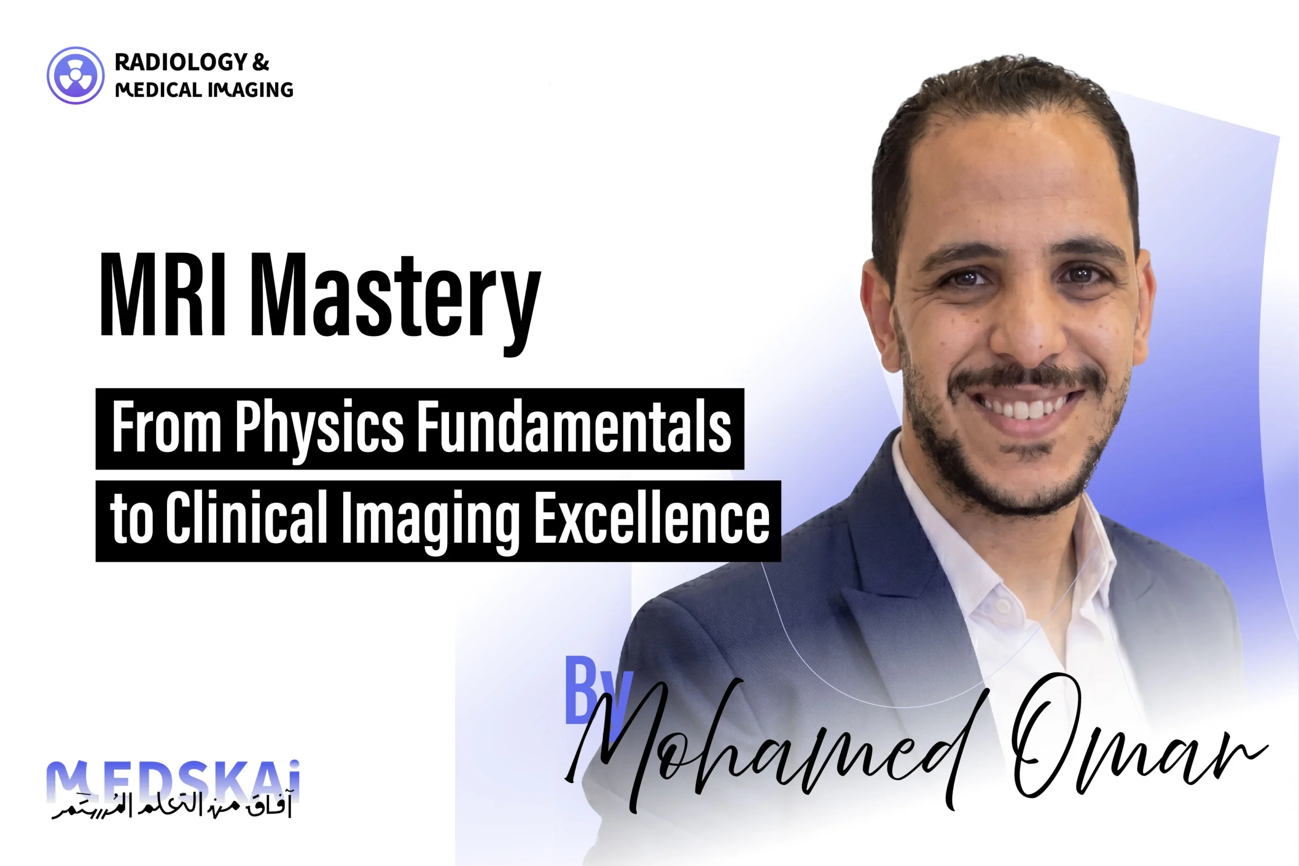
Radiology and Medical Imaging Specialist with over 5 years of clinical and academic experience in diagnostic imaging and healthcare education.Currently working as academic staff, contributing to the delivery of practical and theoretical education in radiology, anatomy, physiology, and imaging processing.Actively involved in curriculum development, academic quality preparation, and student assessment.In parallel, I work as a Radiology Manager at Salamtak Scan Center, overseeing daily operations, quality assurance, staff coordination, and medical reporting processes across multiple imaging modalities.My clinical experience includes hands-on work in CT, MRI, ultrasound, and radiographic imaging, with strong knowledge in radiation protection, image processing, and diagnostic protocols.I am passionate about academic development, medical research, and advancing radiology education to prepare highly qualified graduates for the healthcare market.Open to academic collaborations, research opportunities, and professional networking in radiology and medical imaging.

Community Members Only
Join our community to unlock discussions, share knowledge and connect with members.
Join Discussions
Engage in meaningful conversations
Connect
Build relationships with members
Exclusive Access
Access member-only content

")